https://news.unl.edu/newsrooms/today/article/nine-faculty-earn-named-professorships/
Willa Cather/Charles Bessey professors
Three faculty members were named Willa Cather/Charles Besseyprofessors. The professorship was established in 2001 to recognize faculty members with the rank of professor who have established exceptional records of distinguished scholarship or creative activity.
Qingsheng Li
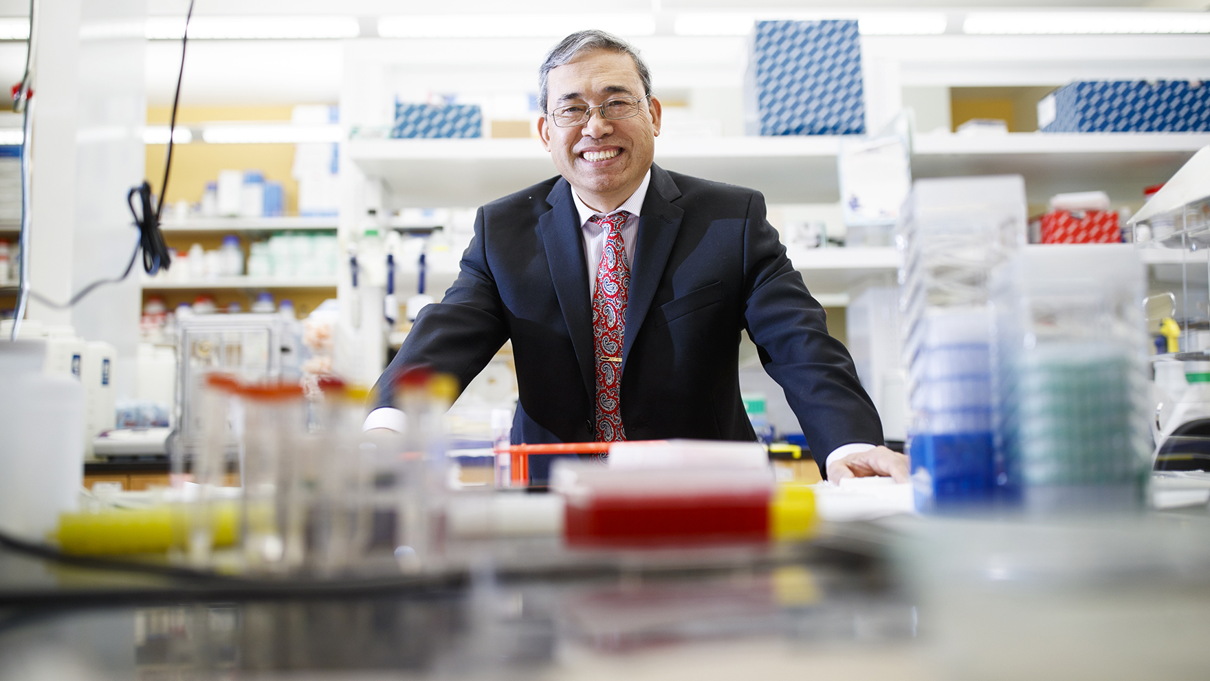
Qingsheng Li will be Willa Cather professor in biological sciences.
Li conducts internationally recognized research on pathogenesis and prevention of viral diseases with an emphasis on human immunodeficiency virus. He has published over 100 peer-reviewed publications, including one in Science, two in Nature, and three in Proceedings of the National Academy of Sciences, and has two patents. Li is currently funded by multiple National Institutes of Health grants as either principal investigator or co-principal investigator and he has received $14 million in external research funds. His contributions to classroom teaching focus on two high-demand, high-enrollment courses with challenging assignments: Human Physiology and Immunology. He is a long serving member of the university's Research Council, Biotech Advisory Board, and Institutional Biosafety Committee. Additionally, Li has served as an NIH panel reviewer and is a standing section member for the HIV Vaccine Development Study Section. He is on the editorial boards of more than a dozen journals and is editor-in-chief of the Journal of Mucosal Immunology Research.
https://news.unl.edu/newsrooms/today/article/wiebe-expands-study-of-poxviruses-immune-function/
Wiebe expands study of poxviruses, immune function
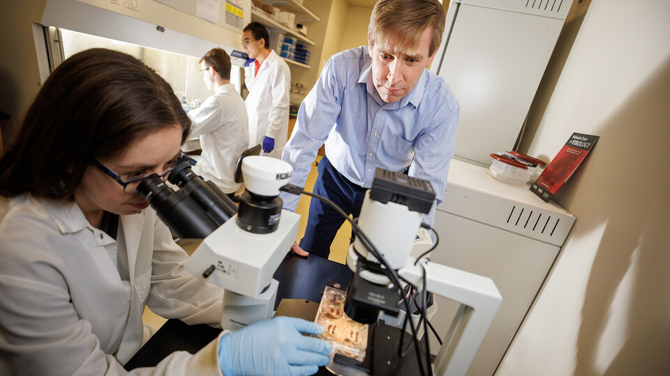
With new funding from the National Institutes of Health, a Husker virologist will advance his work exploring how poxviruses hijack our cellular circuitry during infection, evading host defenses and tuning the environment for unfettered replication.
Matthew Wiebe, an expert in poxvirus-host interactions, recently received a five-year, $2.2 million grant from NIH’s National Institute of Allergy and Infectious Diseases to study how poxviruses capture genes from the host cell, then deploy them to manipulate and disrupt cell signaling pathways during infection. He is particularly interested in how and why poxviruses have evolved to mirror many of the genes and proteins found in host cells.
“One of the ways viruses appear to tap into our cellular circuitry and manipulate it is by having certain genes and proteins that are very similar to ours – but then altering them,” said Wiebe, professor of veterinary and biomedical sciences. “It’s our hypothesis that by understanding why the viruses have made these copies and what they are using the proteins for, we can better understand ourselves, and potentially key weaknesses in our immune system.”
Along with furthering understanding on cellular biology and immune function, findings from the research could identify strategies for fighting viruses and other diseases.
The poxviruses – best known for the variola virus, which causes smallpox – are fertile ground for studying this mimicry. DNA sequencing has revealed close resemblance between viral genes and human cellular genes; closer, even, than the similarities between members of some human gene families.
With the new funding, Wiebe will conduct proteomics and interactome studies on the vaccinia virus to study these “copies” in more detail, exploring how the virus uses the duplicated structures and which cellular signaling pathways they’re involved in. He’ll partner with the university’s Metabolomics and Proteomics Core Facility for some of this work.
Wiebe’s approach is unique in the virology field for its focus on the signaling pathways of kinases and their close cousin, pseudokinases. Kinases are enzymes that catalyze reactions that turn other cellular molecules on and off. Pseudokinases have the same shape and most of the same amino acid pairs as kinases, but they’re missing a few of the critical components driving enzyme activity, rendering them inactive. Wiebe aims to shed light on the purpose of these inactive enzymes in the virus-host interplay.
“Biology doesn’t keep things around, especially in viruses, that have no function,” he said. “So what in the world are these for?”
His hypothesis is that they have multiple roles. Some may regulate traffic, directing other proteins to particular locations in a cell. Some may attach to enzymes, changing their shape and function. And others are likely negative regulators, serving as a brake pedal that slows the pace of certain cellular processes.
Wiebe has been a leader in exploring this third possibility; specifically, his team has demonstrated how the viral pseudokinase B12 inhibits vaccinia replication. Wiebe’s team will extend this trajectory, studying the sometimes counterintuitive ways in which pseudokinases govern viral activity.
Another area of Wiebe’s research is focused on the role of a protein that sits on the frontline of the virus-host battle. BAF – which stands for barrier to autointegration factor – is a tool that either side can harness to its advantage.
The host cell can deploy BAF to thwart viral replication, which it accomplishes by binding to and compacting the viral DNA, making it unreadable. On the other hand, the vaccinia virus can use its B1 kinase to cripple BAF’s DNA-binding ability, making it incapable of stopping proliferation.
Wiebe will advance his work to understand this tug-of-war over BAF. He’ll also investigate other proteins that, like BAF, are capable of playing for either side. These include histones and a protein complex called HUSH, or human silencing hub, a potentially important player in viral expression.
“BAF is the tip of the iceberg,” Wiebe said. “There are many other pathways that are also at the frontline, dictating whether the advantage is shifted to the virus or the cell. Both are fighting over who manipulates certain pathways.”
Revealing the mechanics of these pathways may be relevant to understanding the development of other diseases, such as cancer, infertility, autoimmunity and neurodegeneration. He hopes his research might open the door to new strategies for tackling these problems.
But getting to that point requires embracing the unknown, Wiebe said. That’s why he studies such a broad range of proteins and genes and is excited about novel findings, even when they defy the conventional picture of viral behavior.
“I’m at a stage in my career where I’m trying to embrace confusion, and move toward it,” he said. “There are pros and cons of different genes, and depending on the context, we may detect either one of them. That doesn’t mean it’s not a step forward – it’s another piece of the puzzle and could lead us in a completely novel direction.”
Wiebe’s current team includes postdoctoral researcher Alexandria Krueger and research technicians Zhigang Wang and Ethan Ramsey.
NIH R01 grants are the agency’s original and oldest grant mechanism. They provide support for health-related research and development based on the mission of the NIH.
New vaccine platform could ease development, delivery of virus-fighters
To many, EV stands for “electric vehicle.” To researchers at Harvard University and the University of Nebraska–Lincoln, it’s shorthand for another vehicle — this one nanoscopic — that might help streamline the development and delivery of vaccines worldwide.
By repurposing one of the human body’s natural cargo transports, a Harvard-Nebraska team has developed a vaccine platform that could curb certain engineering challenges, storage demands and side effects of vaccines that combat HIV epidemics and the COVID-19 pandemic. The team’s platform also showed promise in early trials with mice, ramping up antibody production when pitted against HIV and improving survival rates in the face of influenza.
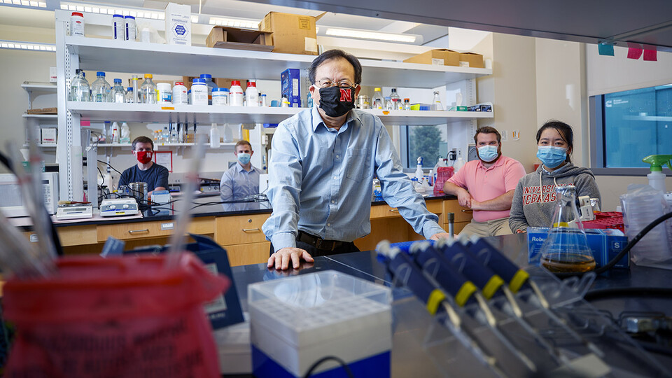
“We believe that this delivery system can enhance the immune response, particularly by inducing antibodies against viral infection,” said Shi-Hua Xiang, associate professor of veterinary medicine and biomedical sciences at Nebraska.
Historically, vaccines have consisted of weakened or inactivated viruses that are recognized by the human immune system, which responds with antibodies and cellular reinforcements that can remember and later fend off infectious forms of those viruses. For reasons of safety and effectiveness, though, many vaccines now instead consist of the lockpicking proteins that coat the surfaces of viruses — so-called antigens that the immune system recognizes as a threat to infiltrate cells.
But to stimulate recognition and immunity, the surface-level antigens in vaccines have to adopt the same three-dimensional poses they strike on viruses themselves. Unfortunately for vaccinologists, coaxing antigens into their native configurations can prove a difficult task. The genetic material of HIV, for instance, is enclosed in an envelope that also stabilizes one of the key proteins protruding from it. Persuading the protein to keep that configuration when uprooted from its viral foundation can mean pairing it with a similar surface or even splicing certain genes into its DNA, a feat that costs time and money.
Across 20-plus years of working on HIV vaccines, Xiang had often engaged in those feats of protein engineering himself. Then, in September 2017, Harvard’s Quan Lu visited Lincoln to give a seminar on a new type of extracellular vesicle: a nanoscopic particle, encased in a double layer of lipids, that ferries nucleic acids and other freight around the body. Xiang, who attended Lu’s talk, knew that lipid bilayers form the membranes of not just EVs and cells, but many viruses, too. The budding process that generated Lu’s EVs also resembled that of viruses. And EVs were roughly the same size as their viral counterparts. Those similarities got Xiang thinking.
“My thought,” he said, “was that maybe we could collaborate on a study.”
Alongside some colleagues, Xiang and Lu were soon looking into whether the latter’s new EV might recruit viral antigens to its surface, lock in their poses, and ultimately stimulate immune responses to them. The researchers started by introducing the EV to an influenza protein that, because of its presence across many different flu strains, has become the subject of the search for a universal flu vaccine. One particular domain of the EV not only drew in and fused with the flu antigen, it also helped promote the budding of more EVs than would otherwise be expected.
Encouraged, the team put its resulting EV to the test in mice. While mice facing the flu without a vaccine survived less than 30% of the time, those given three doses of the EV-based vaccine survived in 60-70% of cases. The vaccine was stimulating high levels of antibodies, the team found, with the antibodies then binding to and neutralizing the flu virus. When the researchers swapped out the flu antigen for an HIV protein, they again saw promising production of neutralizing antibodies in the serum of mice immunized with the vaccine.
Xiang said the platform may boast a couple of advantages even over mRNA vaccines — those that work by instructing cells to churn out antigens and have proven their worth against the SARS-CoV-2 virus responsible for COVID-19. Compared with the mRNA vaccines, which are shipped frozen and can be prone to breaking down over time, an EV-based vaccine will offer more stability and should remain viable at higher temperatures, he said.
Those advantages, Xiang said, could eventually push the EV platform to the forefront of vaccine design, production and delivery.
The team received support from the National Institutes of Health and reported its findings in the journal Science Advances. Lu and Xiang authored the study with Joshua Wiggins, a doctoral student at Nebraska, and Harvard University’s Sengjin Choi, Zhiping Yang, Qiyu Wang, Zhi Qiao and Maoyun Sun.
Eating viruses can power growth, reproduction of microorganism
Over a single day, in the placid waters of a pond, a million virus particles might enter a single-celled organism known for the minuscule hairs, or cilia, that propel it through those waters.
Over the last three years, the University of Nebraska–Lincoln’s John DeLong has been diving into a potential tide-turning secret: Those virus particles are not just a source of infection, they’re also nutrition.
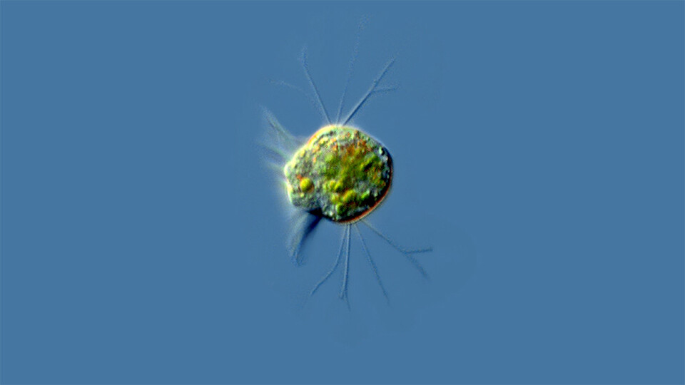
In a turnabout worthy of Pac-Man, DeLong and his colleagues have found that a species of Halteria — microscopic ciliates that populate freshwater worldwide — can eat huge numbers of infectious chloroviruses that share their aquatic habitat. For the first time, the team’s lab experiments have also shown that a virus-only diet, which the team calls “virovory,” is enough to fuel the physiological growth and even population growth of an organism.
Chloroviruses, a career-defining discovery by Nebraska’s James Van Etten, are known to infect microscopic green algae. Eventually, the invading chloroviruses burst their single-celled hosts like balloons, spilling carbon and other life-sustaining elements into the open water. That carbon, which might have gone to predators of the tiny creatures, instead gets vacuumed up by other microorganisms — a grim recycling program in miniature and, seemingly, in perpetuity.

“That’s really just keeping carbon down in this sort of microbial soup layer, keeping grazers from taking energy up the food chain,” said DeLong, associate professor of biological sciences at Nebraska.
But if ciliates are having those same viruses for dinner, then virovory could be counterbalancing the carbon recycling that the viruses are known to perpetuate. It’s possible, DeLong said, that virovory is aiding and abetting carbon’s escape from the dregs of the food chain, granting it an upward mobility that viruses otherwise suppress.
“If you multiply a crude estimate of how many viruses there are, how many ciliates there are and how much water there is, it comes out to this massive amount of energy movement (up the food chain),” said DeLong, who estimated that ciliates in a small pond might eat 10 trillion viruses a day. “If this is happening at the scale that we think it could be, it should completely change our view on global carbon cycling.”
‘Nobody noticed it’
DeLong was already familiar with the ways that chloroviruses can entangle themselves in a food web. In 2016, the ecologist partnered with Van Etten and virologist David Dunigan to show that chloroviruses gain access to algae, which are normally encased in a genus of ciliates called Paramecia, only when tiny crustaceans eat the Paramecia and excrete the newly exposed algae.
That finding put DeLong in “a different headspace” when it came to thinking about and studying viruses. Given the sheer abundance of viruses and microorganisms in the water, he figured it was inevitable that — even setting aside infection — the former would sometimes wind up inside the latter.
“It seemed obvious that everything’s got to be getting viruses in their mouths all the time,” he said. “It seemed like it had to be happening, because there’s just so much of it in the water.”
So DeLong dove into the research literature, intent on surfacing with any studies on aquatic organisms eating viruses and, ideally, what happened when they did. He emerged with precious little. One study, from the 1980s, had reported that single-celled protists were capable of consuming viruses, but delved no further. A handful of papers from Switzerland later showed that protists seemed to be removing viruses from wastewater.
“And that was it,” DeLong said.
There was nothing about the potential consequences to the microorganisms themselves, let alone the food webs or ecosystems they belonged to. That surprised DeLong, who knew that viruses were built not only on carbon but other elemental cornerstones of life, too. They were, at least hypothetically, anything but junk food.
“They’re made up of really good stuff: nucleic acids, a lot of nitrogen and phosphorous,” he said. “Everything should want to eat them.
“So many things will eat anything they can get ahold of. Surely something would have learned how to eat these really good raw materials.”
As an ecologist who spends much of his time using math to describe predator-prey dynamics, DeLong wasn’t entirely sure how to go about investigating his hypothesis. Ultimately, he decided to keep it simple. First, he’d need some volunteers. He drove out to a nearby pond and collected samples of the water. Back at his lab, he corralled all of the microorganisms he could manage, regardless of the species, into drops of the water. Finally, he added generous portions of chlorovirus.
After 24 hours, DeLong would search the drops for a sign that any species seemed to be enjoying the company of the chlorovirus — that even one species was treating the virus less like a threat than a snack. In Halteria, he found it.
“At first, it was just a suggestion that there were more of them,” DeLong said of the ciliates. “But then they were big enough that I could actually grab some with a pipette tip, put them in a clean drop, and be able to count them.”
The number of chloroviruses was plummeting by as much as 100-fold in just two days. The population of Halteria, with nothing to eat but the virus, was growing an average of about 15 times larger over that same timespan. Halteria deprived of the chlorovirus, meanwhile, wasn’t growing at all.
To confirm that the Halteria was actually consuming the virus, the team tagged some of the chlorovirus DNA with a fluorescent green dye before introducing the virus to the ciliates. Sure enough, the ciliate equivalent of a stomach, its vacuole, was soon glowing green.
It was unmistakable: The ciliates were eating the virus. And that virus was sustaining them.
“I was calling up my co-authors: ‘They grew! We did it!’” DeLong said of the findings, now detailed in the journal Proceedings of the National Academy of Sciences. “I’m thrilled to be able to see something so fundamental for the first time.”
DeLong wasn’t done. The mathematical side of him wondered whether this particular predator-prey dynamic, as strange as it seemed, might share commonalities with the more pedestrian pairings he was accustomed to studying.
He started by charting the decline of the chlorovirus against the growth of the Halteria. That relationship, DeLong found, generally fits with those ecologists have observed among other microscopic hunters and their hunted. The Halteria also converted about 17% of the consumed chlorovirus mass into new mass of its own, right in line with percentages seen when Paramecia eat bacteria and millimeter-long crustaceans eat algae. Even the rate at which ciliates preyed on the virus, and the roughly 10,000-fold disparity in their sizes, match up with other aquatic case studies.
“I was motivated to determine whether or not this was weird, or whether it fit,” DeLong said. “This is not weird. It’s just that nobody noticed it.”
DeLong and his colleagues have since identified other ciliates that, like Halteria, can thrive by dining on viruses alone. The more they uncover, the more likely it seems that virovory could be occurring in the wild. It’s a prospect that fills the ecologist’s head with questions: How might it shape the structure of food webs? The evolution and diversity of species within them? Their resilience in the face of extinctions?
Again, though, he’s opted to keep it simple. As soon as Nebraska’s winter relents, DeLong will head back to the pond.
“Now,” he said, “we have to go find out if this is true in nature.”